/laglivlab-illustrasjon-english-1000px.png)
1 Project Planning
-
Brainstorming and wild ideas are invited. Everyone must search online, in (popular) scientific journals and newspapers to find life science technology and research they find exciting. To give you some ideas, search online with some of these keywords:
- Cell culture on a chip
- 3D cell cultures
- Bioimpedance on cells
- Acoustic/electrical rotation of cells
- Soft robotics: muscle cells in robots
-
The supervisors will explain the limitations we have due to available equipment, cost, and time.
- The students and supervisors will discuss the remaining, possible project ideas and decide to focus on one or two projects.
-
Interdisciplinary project teams will form around their project. The biological, chemical, medical, physical, electronic, and data processing aspects of the project
- are mapped out by those with the best expertise.
- are taught to the others on the team.
-
The team decides on the goal of the project, what type of cells are needed, how to influence the cells to achieve what is desired, and how to measure the changes.
- Plans are made for sub-projects and who will participate in them.
2 Sub-projects
In the next phase, it may be natural for the team to split up and work in parallel on sub-projects they are most interested in or have the best prerequisites for. But all the time, the different sub-projects must communicate so that they fit together in the end.
2.1 Selection and preparation of cell cultures, nutrient fluids, and "medicines"
In collaboration with our partners at the Hybrid Technology Hub and other biological and medical research groups, available cell lines that these groups have at their disposal will be selected. The cell lines will be thawed and cell culture will be started and maintained by the students. The necessary nutrient fluids and medicines will be purchased or mixed, and protocols for different parts of the process in traditional cell cultivation will be established. Protocols for corresponding processes in microfluidics will be planned. Any new fluorescence markers will be included and tested.
2.2 Design of microfluidics and electrodes
This is done on the students' computers. It can be anything from simple drawings to drawings based on simulations of fluid flow or electric fields. The drawings are printed on special transparencies used in photolithography.
2.3 Simulation
If the team chooses to do so, they can perform simulations of parts of the processes included in the project.
2.4 Microfabrication
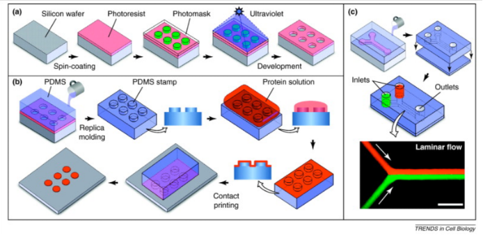
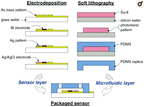
The students themselves perform photolithography and microfabrication of microfluidics and electrodes. See figures 1 and 2.
Microfabrication of microfluidics:
2.5 Measurement systems and influencing the cells
2.5.1 Microscopy
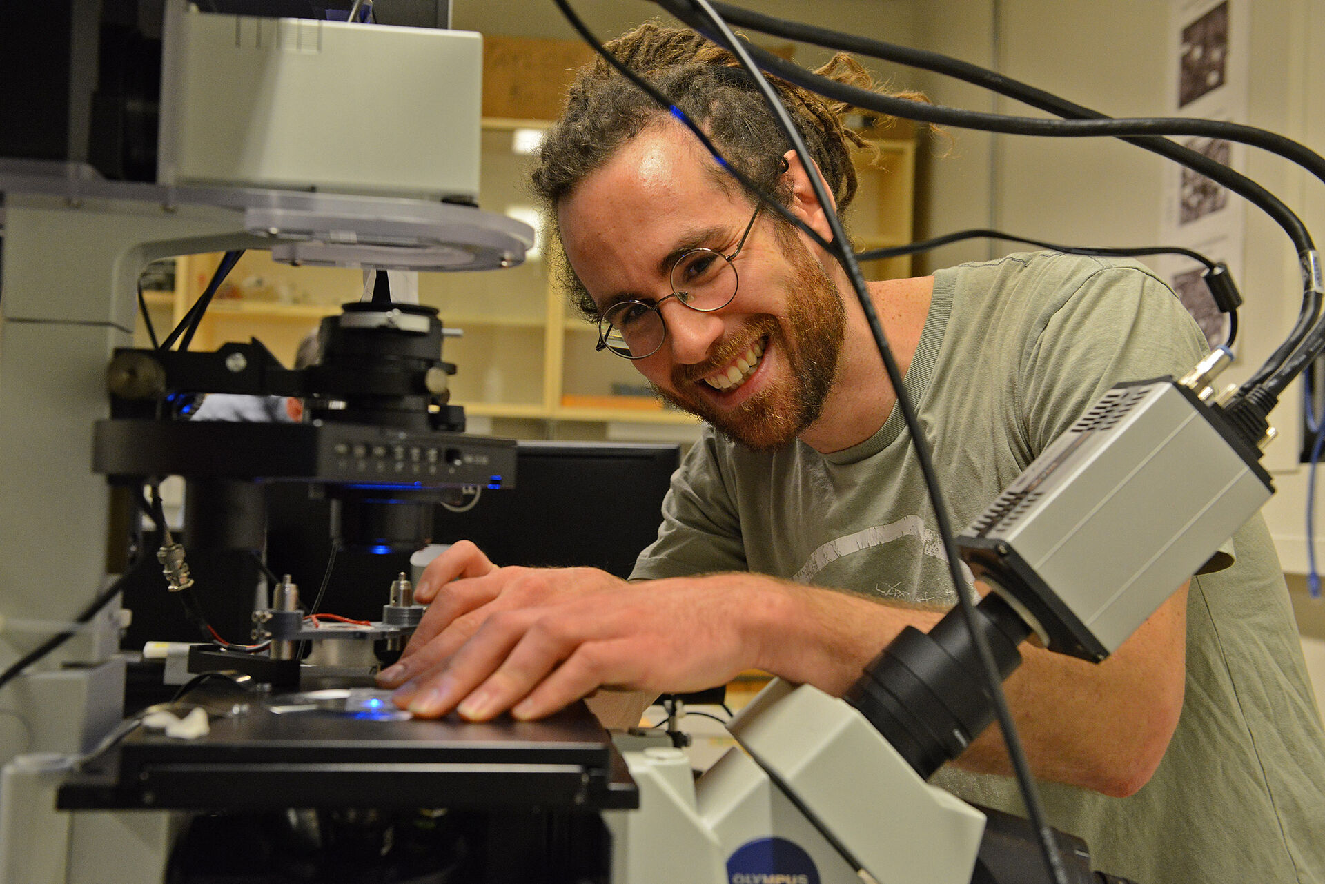
Fluorescence microscopy is a basic technique in modern biology. Biology and medical students have probably used it. In addition, we can use other optical measurement methods and custom-build microscopes. Physicists and mathematicians have a good background for analyzing the images in a quantitative way.
2.5.2 Electrical: Bioimpedance and electrochemistry
Electrodes can either be used to transfer electrons from substances in the fluids (electrochemistry) or to measure dielectric changes between the electrodes (impedance measurements). Electrochemistry can be used to measure concentrations of specific substances in the fluid, also in microfluidics. Bioimpedance measurements on cell cultures in microfluidics are brand new.
Example of microfabrication of electrodes in microfluidics:
2.5.3 Ultrasound and other techniques
We have equipment and experience in influencing cells with ultrasound, pressure, and fluid flow.
3 Testing the system and experiments
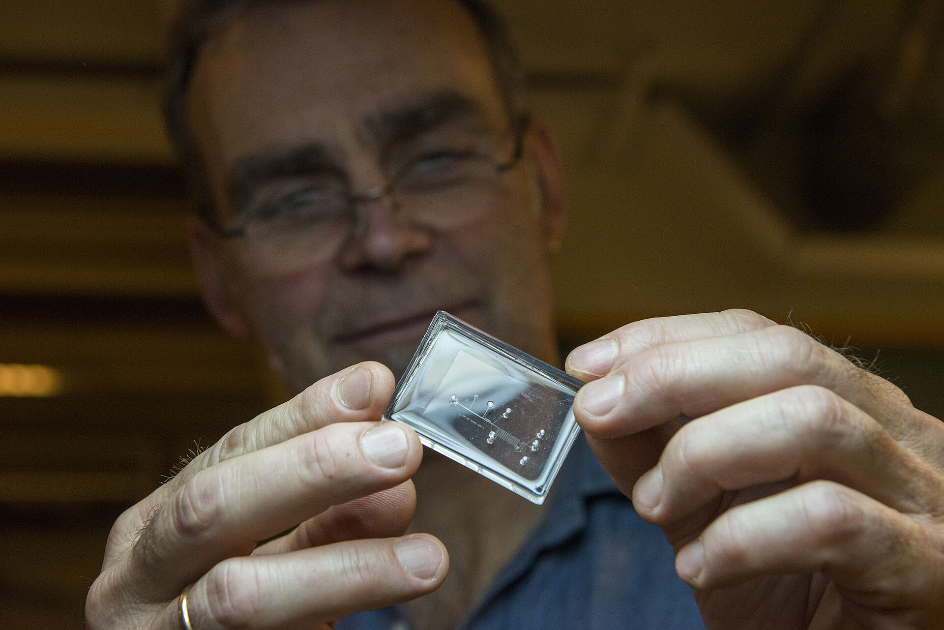
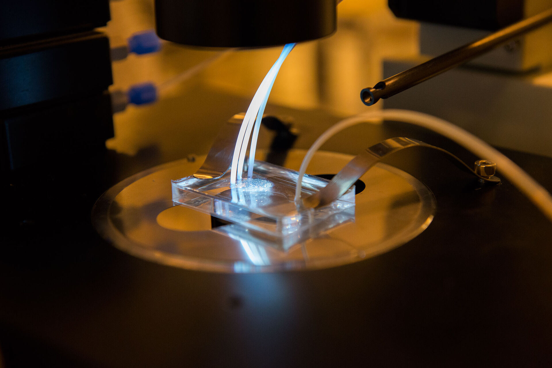
At this stage, everyone on the team will work together to assemble the different parts of the project. The microfluidic network with electrodes is connected to the fluid control system and the measurement electronics, and it is placed in the microscope to monitor and measure everything that happens. Fluids and cells are released into the network and the experiments begin...
4 Analysis of experiments and comparison with simulation
At this stage, it is once again important to have everyone involved. Biologists and medics understand the biological changes to be followed. Together with the other realists, this is converted into quantitative measures that can be obtained from the images and electronic measurements.
5 Scientific publication or a patent application?
It's hard to say how far the project will get. Whether the goal is scientific or technological, the team must document and summarize what they have done and learned during the project. Even if it does not become a scientific publication or a patent application after one semester, it may become one from the continuation. And the joint report is in any case good to show to future supervisors and employers.