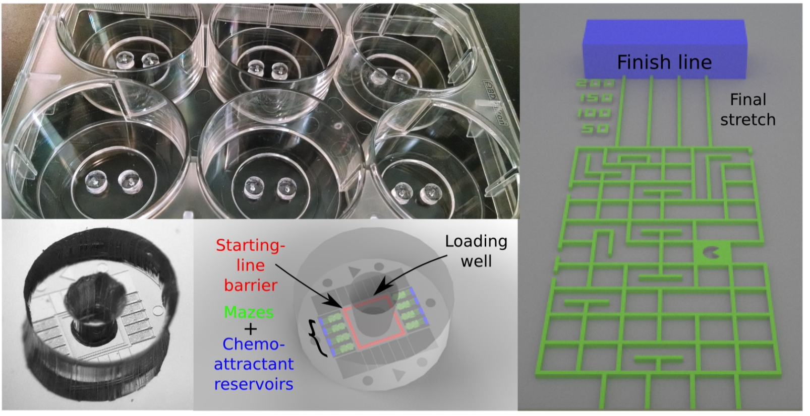
In 2014 teams from around the world participated with their favourite cells in the Dicty World Races. The cells were attracted by a chemical (a so-called chemoattractant) that was most concentrated at the finish line. But to reach the finish line they had to find a path through a maze of micro-tunnels. The maze with all the cells were imaged to track the cell motion and analyse it. The video of cells in the maze gives you an impression of how one cell type moved.
There are many fun, artistic and scientifically interesting variations to such a cell race through a maze. Make your own fun maze? Let the cells find their way through the PoreLab maze that is the result of a fascinating self-organised process? Let the maze be coloured by the cell speed or the number of cells passing by? Let tissue-forming epithelial cells compete with immune cells? Try to make numerical models of the cell motion?
-
Science:
- Do individual cells move faster than a tissue?
- Do cells navigate and move faster in tunnels or on tracks?
- Can we model the cell motion numerically?
-
Fun:
- Competing teams
- Department of silly walks
-
Beauty:
- Construct beautiful mazes
- Let cell's motion draw pictures, colour the maze, ....
-
Learning:
- Photolithography
- Microfluidics
- Image analysis
- Cell culture
- Modelling
Plan for autumn 2025
Designing photomasks for labyrinth and other fibronectin and microfluidic designs
Starting Sep 4th we will Install AutoCAD and draw designs on our computers following these instructions (under subheading "Designing Microfluidics mask"). Bring your own laptop!!
Cells attach to surfaces (very few can swim). We use thin glass as surfaces for cell experiments because then we can easily image the cells. The glass surface needs to be "adhesive" in the sense that the cell can connect it's adhesion molecules to the surface. We will learn methods to cover the glass surface with adhesion molecules. There are two "engineering methods" of restricting where cells can move. Both methods can be patterned with micrometer precision by photolithography:
- Microfluidic channels: Then cells are moving inside a channel with liquid and hard side walls restrict them from going anywhere else..
- Adhesive substrate: Cells will only go where there are adhesive molecules and all other areas of the surface will be restricted by a non-adhesive layer. In our first round of CellMazeRace we will use this method because it is very easy to seed the pattern and keep the cells alive.
We can use exactly the same mask patterns for both methods, the only difference is that black and transparent has to be inverted. This means that if you want to make photomask patterns for other purposes (microfluidics or fibronectin) you can make them now. When the designs are finished we will send them to a company that will print them on fused silica. When we get the physical mask back we are ready to do the photolithography.
Design questions:
- How wide and long should the paths be?
- How do we avoid cells attaching on top of a pattern, that is, how do we give them a starting area?
Keeping cells alive
Starting Sep 11th we will go to the cell lab and learn the steps necessary to keep a cell culture alive. We will start with MDCK cells and the protocol is describe here.
Monitoring of cell motion and development
Starting Sep 11th we will also start a first experiment of cells on a adhesion pattern. We will use the HoloMonitor microscope to make timelapse videos of the cells.
Analysing cell motion and development
Starting Sep 18th we will view and start analyzing the timelapse videos of the cells moving on the adhesion pattern.
Deep UV lithography for fibronectin patterns
Starting Sep 25th we will use this procedure to transfer the pattern from the photomask to an adhesive pattern of fibronectin molecules
CellMazeRace
When all pieces have been put together.