Project ideas for LagLivLab
Aug 28, 2025
/laglivlab-illustrasjon-english-1000px.png)
Dynamic patterning of chlamydomonas reinhardtii (TC)
This is inspired by the work of the artist Lia Giraud (http://www.liagiraud.com/centre-pompidou/ - in French).
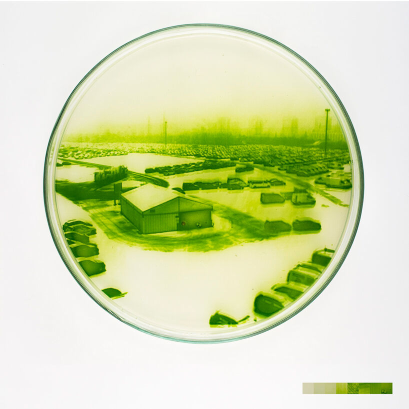 |
Figure 1: Example of reproduced photography with micro-algae. Copyright: Lia Giraud. |
Chlamydomonas reinhardtii is an unicellular algae that is capable of locomotion in a liquid media (micro-swimmer) which exhibits phototaxism: it will preferably direct its motion toward a light-source. This capacity has been used by researcher to create directed motion of micro-swimmer assembly (cf. work by Rafai et al.), but also by artists in order to create "biological pictures": illuminated thought a pattern, these algae will concentrate more in the brighter areas, reproducing the initial pattern.
We propose to explore both the scientific and artistic point of view with a similar setup. In Giraud's work, the mobility of the algae was removed once the image was formed (agar-agar?), but it would also be interesting to see the potential of a similar system to create dynamic images, by changing the underlying pattern for instance with a video-projector. This brings interesting scientific questions like: How long should a pattern be present for the algae to form a convincing pattern? Once produced, what is the remanence of this pattern when the illumination is removed.
Once the system is mastered, one can use it for studying more basic scientific questions, for instance by using continuously changing patterns to try to induce global micro-swimmers motion.
CellMazeRace
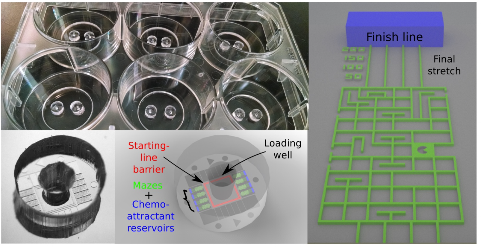 |
Figure 2: Dicty World Race in 2014: Pictures and sketches of the microfluidic devices and the racecourse. Each well of a 6-well plate contained two devices and each device contained 8 maze racecourses |
In 2014 teams from around the world participated with their favourite cells in the Dicty World Races. The cells were attracted by a chemical (a so-called chemoattractant) that was most concentrated at the finish line. But to reach the finish line they had to find a path through a maze of micro-tunnels. The maze with all the cells were imaged to track the cell motion and analyse it. A video of cells in the maze gives you an impression of how one cell type moved.
There are many fun, artistic and scientifically interesting variations to such a cell race through a maze. Make your own fun maze? Let the cells find their way through the PoreLab maze that is the result of a fascinating self-organised process? Let the maze be coloured by the cell speed or the number of cells passing by? Let tissue-forming epithelial cells compete with immune cells? Try to make numerical models of the cell motion?
- Science:
- Do individual cells move faster than a tissue?
- Do cells navigate and move faster in tunnels or on tracks?
- Can we model the cell motion numerically?
- Fun:
- Competing teams
- Department of silly walks
- Beauty:
- Construct beautiful mazes
- Let cell's motion draw pictures, colour the maze, ....
- Learning:
- Photolithography
- Microfluidics
- Image analysis
- Cell culture
- Modelling
Skin battery
Electric potential differences can easily be measure with a voltmeter between different skin sites on the human body. A voltage of 20 – 30 mV can e.g. typically be found between the skin on the volar part of the underarm and the palm of the hand. Relatively little is known about the source of these potentials, but mechanisms such as Donnan potentials or streaming potentials (due to sweat secretion) are possible candidates. This project provides possibilities for both theoretical work and experimental work. More to come ...
Finding the source of the potentials
Skin is a layered structure with solid materials and electrolytes. More to come ...
Electrodermal activity
By measuring the potential between the underarm and the palm of the hand as a function of time, one will see that the potential fluctuates with the degree of psychological stress. What is the mechanism behind and what can this be used for? There is scientific literature that describes some application areas for such measurements. More to come ...
Can energy be harvested from these sources?
How much power can be drawn from these bodily voltage sources? Experiments can be carried out where the voltage sources are loaded by a parallel (variable) resistor while the voltage is measured. From these measurements the maximum power can be decided. Can this energy be harvested? Can the students construct an electronic circuit for this purpose? More to come ...
Electrorotation
The electrorotation project has been running for several years and the results are close to ready to be published. Do you want to participaqte in the last leg of the journey?
Microfluidics
/labonachip-laglivlab-1076x718.jpeg) |
Figure 3: Lab-on-chip system |
Microfluidics, a cutting-edge technology that manipulates tiny volumes of fluids at the microscale, is revolutionizing the life sciences. By controlling and analyzing small amounts of liquid—often in the range of microliters to nanoliters—microfluidics enables researchers to conduct intricate experiments with unprecedented precision and efficiency.
There have been several microfluidics group projects at LagLivLab . We are now open for new projects. Here is a short list of areas in life science where microfluidics is used:
- Diagnostics:
- Point-of-Care Testing: Rapid tests for diseases like HIV, malaria, and COVID-19.
- Blood glucose monitoring devices for diabetes management.
- Microfluidic-based assays for detecting specific biomarkers in bodily fluids.
- Genomics:
- High-throughput DNA sequencing: Utilizing microfluidic chips to streamline the sequencing process.
- PCR amplification of DNA samples: Reducing time and materials required for amplification.
- Single cell RNA sequencing: Enabling analysis of gene expression at the single-cell level.
- Proteomics:
- Protein separation and analysis via microfluidic electrophoresis and chromatography.
- Enzyme activity assays to study biochemical reactions in small volumes.
- Antibody screening and development in microfluidic systems.
- Cell Biology:
- Cell culture in microenvironments: Mimicking in vivo conditions for more accurate biological studies.
- Single-cell analysis: Studying cellular heterogeneity by isolating and analyzing individual cells.
- Drug response assays: Testing the effects of pharmaceuticals on specific cell types.
- Tissue Engineering:
- Microfluidic scaffolds for cell growth and tissue development.
- Organ-on-a-chip models: Simulating the physiological responses of human organs for drug testing or disease modeling.
- Environmental Monitoring:
- Detection of pollutants and toxins in water and air samples.
- Microfluidic biosensors for monitoring microbial activity in environmental samples.
- Synthetic Biology:
- Engineering and screening of microorganisms for biofuel production or bioremediation.
- Automated assembly of DNA constructs and pathways.
- Biotechnology:
- High-throughput screening of biochemical reactions and metabolic pathways.
- Optimization of fermentation processes in microbial biotechnology.
- Pharmaceutical Development:
- Early-stage screening of drug candidates in miniaturized systems.
- Pharmacokinetics studies: Understanding how drugs are absorbed, distributed, metabolized, and excreted.
- Microbiome Research:
- Study of microbial communities in various environments and their interactions with host organisms.
- Analysis of single microbial cells for genomic and phenotypic characterization.
- Immunology:
- Development of microfluidic immunoassays for detecting antibodies or antigens.
- Studying immune cell interactions under controlled conditions.
- Biophysical Studies:
- Measurement of cellular forces and mechanics using microfluidic devices.
- Analysis of the physical properties of cells such as stiffness, adhesion, etc.