About the project
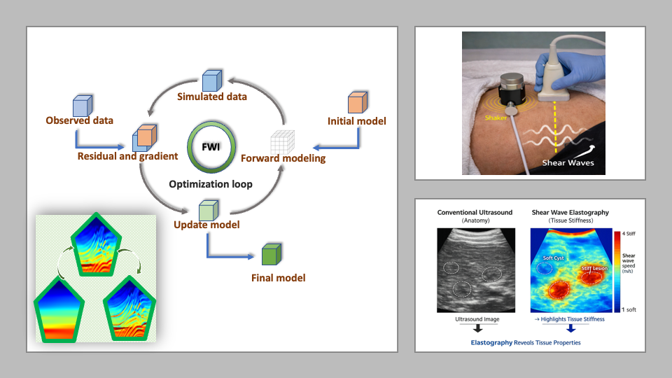
Ultrasound elastography is a non-invasive and cost-effective technique for diagnosing diseases by measuring tissue stiffness. Safer and more portable than magnetic resonance elastography (MRE), it is well-suited for clinics and resource-limited settings. This study focuses specifically on time-harmonic shear waves generated by an external actuator, which offer enhanced tissue illumination and are particularly effective for imaging deeper organs.
In this project, we propose the development of an innovative processing approach based on Full Waveform Inversion (FWI) for time-harmonic shear wave elastography. Known as a powerful tool for seismic imaging and subsurface exploration, FWI has also shown potential in medical imaging, as both fields share similar principles and techniques in signal processing. FWI can be described as a PDE-constrained optimisation method that reconstructs material properties by matching simulated and measured wavefields.
However, while historically constrained by computational burdens, advancements in technology and data science now enable its broader application. We aim also to leverage machine learning approaches, such as neural operator methods, to accelerate the computationally intensive simulations of wavefield propagation and mode conversion. This will contribute to a significant advancement, enhancing both the efficiency and accuracy of the FWI imaging technique.
An illustrative application could involve assessing cardiac health and diagnosing conditions by estimating myocardial stiffness. Validation against state-of-the-art techniques will be carried out using simulation studies, channel data recorded from elasticity phantoms, and in vivo data acquired with the research ultrasound scanners available in the DSB group laboratory at the Department of Informatics.