High frame rate ultrasound (US) imaging, the capability of achieving 3,000 to 10,000 frames per second (fps), has garnered attention for its utility in functional imaging. The concept of high frame rate imaging was introduced to medical ultrasound imaging in the 1970s through parallel receive beamforming. Despite remarkable temporal resolution, practical application of high frame rate ultrasound was delayed for years due to hardware limitations. Over the past two decades, numerous approaches have been developed to enhance the frame rate of ultrasound imaging systems. Today, programmable ultrasound imaging systems have become available, allowing the implementation of transmit-receive sequences that facilitate high frame rate imaging. The imaging mode is particularly suited for assessing the dynamic properties of tissues, such as blood flow and the propagation of transverse waves.
The DSB group has an ongoing research activity in ultrasound elastography imaging. For this we use high frame rate imaging. Our imaging modality is based on the use of plane or divergent waves, as opposed to focused beams which are used for lower frame rate and high quality imaging. Plane and divergent waves are generated by applying a plane or circular delay on the elements of the transducer. In both cases, it is possible to reconstruct an entire image with a single emission. The imaging approach result in reduced spatial resolution and contrast of the images compared to images acquired useing focused beams. A technique that can be used to overcome these limitations is to use multiples plane or diverging waves transmitted at slightly different angles to obtain one image. The price to pay for this is a reduction of the frame rate, i.e. it is possible to trade frame rate for image quality and vice versa.
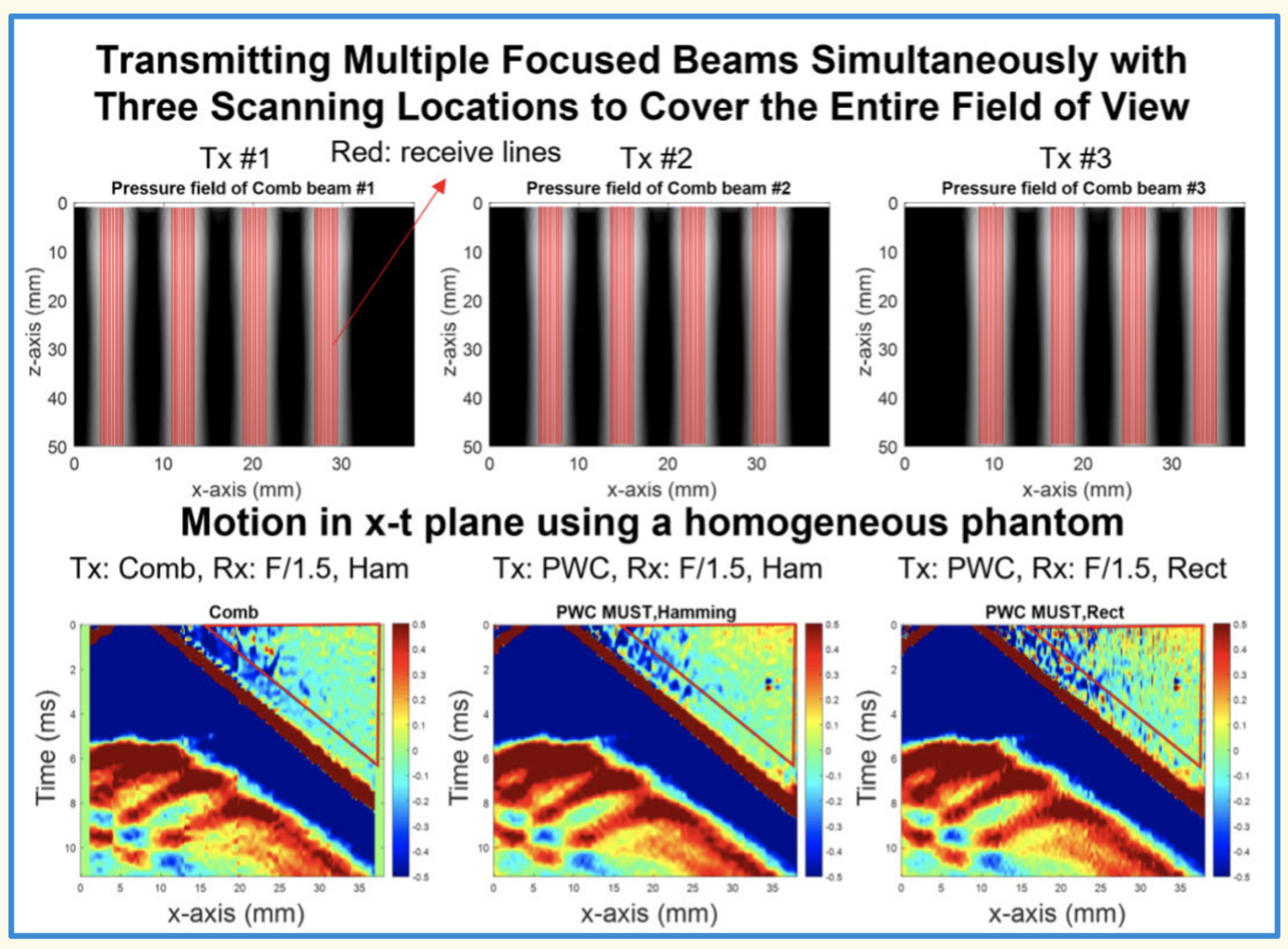
Recently, an alternative acquisition scheme has been introduced: comb beams [1]. It promises high frame rate and increased signal-to-noise ratio compared to plane wave imaging.
In this project, we aim to implement and compare comb beam imaging to plane and diverging wave imaging.
In the project, you will do the following:
- Study the literature on high frame rate imaging
- Implement comb beam imaging in USTB
- Test and compare comb beam, plane, and diverging wave imaging on simulated ultrasound images using Field II
- Implement comb beam imaging on our experimental ultrasound imaging system
Qualifications:
- Excellent MATLAB programming skills
- Signal processing (IN3190/4190, IN3015/4015, IN5450, INF4480)
[1] Lee, Hyoung-Ki, Philip M. Holmes, James F. Greenleaf, and Matthew W. Urban. ‘Comb Detection for Measuring Shear Wave Propagation’. IEEE Transactions on Ultrasonics, Ferroelectrics, and Frequency Control 70, no. 9 (September 2023): 1135–45. https://doi.org/10.1109/TUFFC.2023.3297394.