Cardiac MR elastography (cMRE) is a new imaging modality that produces stiffness images of the heart. The technique is based on the fact that mechanical waves move faster through stiff tissue than soft tissue. By recording the movement of waves in the heart, we can use specialized algorithms to detect wave speed and to there by infer tissue stiffness.
This technique fills a key method gap that has persisted in cardiac imaging for decades and provides a new dimension that could significantly change our understanding of numerous diseases and conditions.
The use of MRE in the heart is still in its infancy, meaning that many questions remain unanswered. Importantly, the effects of differing heart geometries and wave patterns on the stiffness output of the MRE algorithms is largely unknown. This means that researchers cannot trust whether changes to stiffness output comes from true changes to tissue stiffness and wave speed, or from undesired side effects of geometry and wave behavior. In this project the student will answer these questions by creating digital models using in vivo data, where the heart geometry and behavior of waves are used as parameters to influence stiffness output in different MRE algorithms. This will provide answers to how different MRE algorithms respond to non-stiffness changes in tissue, and thereby improve the reliability of our in vivo experimental results.
Plan for the project:
- Establish digital mechanical wave model of the heart with controllable wavelength, wave amplitude, wave attenuation, wave noise, and wave direction.
- Expand model to include several heart geometries, including those associated with common pathophysiological states such as hypertrophy.
- Compare stiffness output of different MRE algorithms to true digital model stiffness, with and without manipulation of parameters established in 1. and 2.
- [If time allows:] Improve established MRE algorithms or implement new ones.
- [If time allows:] Implement wave amplifying or de-noising strategies to improve performance of MRE algorithms both in vivo and in silico.
Necessary competence:
- Strong programming skills
- A background that includes mathematics or physics
Desirable competence:
- Experience in using MATLAB or Python for data analysis
- Experience with image analysis
- Understanding of mechanical wave behavior
- Understanding of viscoelastic physics
- Understanding of Fourier transform or other relevant integral transforms
- Visual communication skills
- Teamwork skills
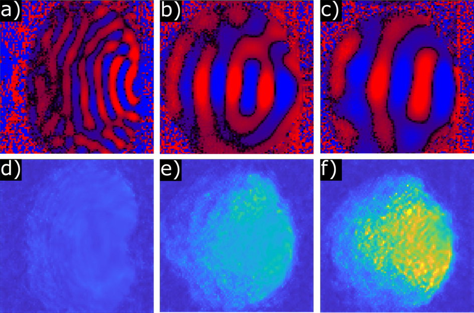
a-c) show MRI phase images that capture mechanical waves travelling through 3 homogenous gel phantoms with increasing stiffness. The waves come in from the right side and propagate towards the left. Noisy areas are outside the gel. The wavelength (and implicitly, wave speed) increases with the stiffness of the gel. d-f) show the resulting stiffness images output by one MRE algorithm. The algorithm correctly detects increasing stiffness yet fails to capture the homogenous stiffness across the gel. The algorithm appears to fail near the edges of the gel and where the wave attenuates.

a) shows a MRI magnitude image of a rat heart. Position of heart wall is marked in green for clarity. b) shows an MRI phase image. Mechanical waves are propagating into the heart from the right. c) Shows the segmented stiffness image. A homogenous, moderate stiffness across the myocardium can be seen, as expected in a healthy heart. But how much does the geometry of the heart and the changes to wave behavior affect the output? That will be answered through this master’s project.