Please come see us for further dialogue if you're interested in working on these interesting research directions.
Examples of such research directions are:
1) Elastography
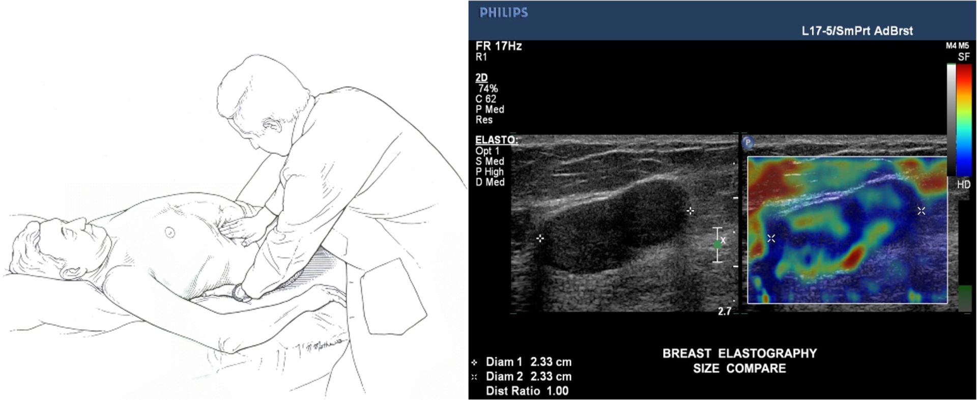
We have several interesting topics to tackle in the domain of mapping tissue stiffness using Time harmonic elastography.
Walker HK, Hall WD, Hurst JW
2) Ultrasound localization microscopy (ULM)
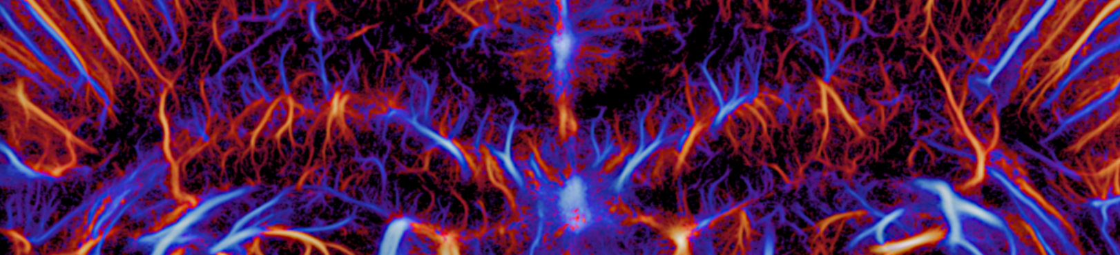
In ULM, microbubble contrast agents are injected in to the bloodstream. By tracking the path of individual bubble, an extremely detailed view of the microvascular structure can be achieved. This has several important applications in, for example, imaging the brain.
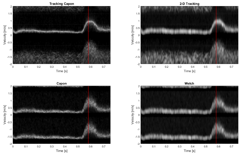
3) Spectral Doppler for blood flow characterization
Ultrasound spectral Doppler is used to assess how blood flow at a specific location changes through time. This information is used for instance for detection of stenotic arteries as well as the degree of stenosis.
4) Image reconstruction and grating lobe suppression
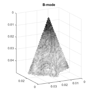
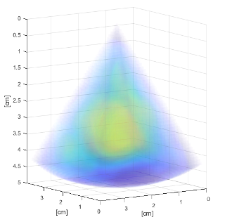
There are several reasons why ultrasound imaging systems often feature sensors that are distributed at a distance which is greater than half the wavelength of the ultrasound pulse. This spatial undersampling generates artifacts called grating lobes, where non-physical "copies" of true structures appear in the image. We will look into methods to bot identify and to reduce such artifacts in the final ultrasound image.
Necessary experience
- To have taken or later take the course in Ultrasound imaging (IN3015/IN4015)
- To have taken or later take the course in Signal processing (IN3190/IN4190)
- To take the course in Array signal processing (IN5450)
- Strong scientific programming skills
Additional beneficial experience
- Wave physics
- Interest in medical technology