About the project
Pollution of soil and ground water is a serious health threat where heavy metals express one of the highest hazards. Cadmium (Cd) is a resilient heavy metal which is among the top six pollutants worldwide. Therefore, understanding and quantifying the retention and sorption mechanisms of cadmium in rocks is of tremendous importance for both risk preparedness and hazardous waste treatment.
We use in-situ neutron imaging of Cd-doped flow experiments in limestone and sandstone samples. While the Fontainebleau sandstone was completely washed and no traces of cadmium remained in the rock, the Indiana limestone has demonstrated a real potential in retaining cadmium due to two different porous network sizes and permeabilities. At the end of the experiment about 30% of the injected cadmium remained in the rock, unevenly distributed in the rock, highlighting some areas of stagnant fluids. It provides a potential new perception of the capture of pollutants where only a fraction of the rock structure contributes to the sorption of heavy metals.
The first year of this project investigated the project feasibility through fast 2D radiographies. Successful results already provided a first publication (Cordonnier et al., 2019) and motivation for advanced techniques. Accordingly have adressed the development of fast 3D tomography with scattering correction in order to quantitatively better image the small variations of our pollutant tracer and localize them in a three dimensional space.
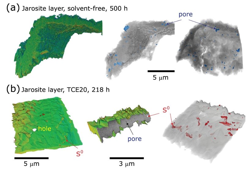
Through a collaboration with Prof. Fang Xia at the Murdoch University in Australia, we have imaged chemical processes occurring during the leaching of ore minerals. We also continued our developments of X-ray imaging processes using the Hades rig and developed several data processing tools to image and predict fracturing processes in rocks.
Financing
The Research Council of Norway (project ARGUS, #272217, 2017-2020).
Cooperation
- The Njord Centre, University of Oslo, Norway
- University Grenoble Alpes, Grenoble, France
- Paul Scherrer Institute, Villigen-PSI, Switzerland
- University of Edinburgh, Edinburgh, UK
- Technische Universiteit Delft, Delft, Netherlands