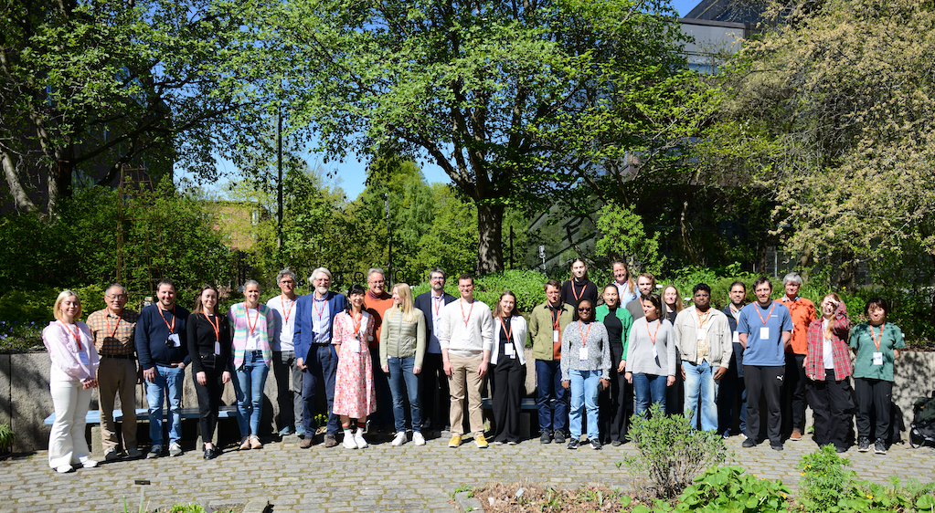
This spring, the Biological Optical Microscope Workshop resumed after being suspended in 2024 due to the organizing team's preparations for the international microscopy conference, BNMI 2024, in Geilo. The conference aimed to encourage collaboration and networking among researchers and facilitate the sharing of facilities among Scandinavian countries. Upon reopening, several students and researchers from across the country eagerly awaited the chance to register.
Intensive laboratory sessions
Alongside theoretical lectures, the workshop's afternoons were filled with hands-on laboratory sessions. On the first day, optical labs were conducted to demonstrate the basic properties of light and microscope components, with content updated annually based on faculty availability, teaching materials, and current trends in microscopy.
Innovative diffraction lab introduction
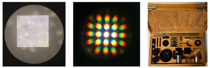
This year, a new diffraction lab was introduced featuring an antique Zeiss diffraction kit, designed by Ernst Abbe in 1965. The kits, recovered from James Pawley’s microscopy workshop in Vancouver, allow for the visualization of enlarged images of different gratings and the back focal plane, presenting microscopy images in frequency space (Fourier transform), commonly utilized in image processing.
Demonstrations provided insights into the relationship between an image and its back focal plane, and how manipulating the back focal plane affects resolution, contrast, and image details. Teaching staff worked collaboratively to restore the kit, with Rainer Heintzmann demonstrating its functionality. Students expressed enthusiasm for the diffraction lab and suggested allocating more time for it in future workshops. These demonstrations fascinated both students and faculty, offering unique experiences at the NorMIC workshop.
Microscope hands-on labs
The afternoons were filled with hands-on microscopy sessions where students rotated through groups among various microscopes, exploring diverse imaging modalities. This aspect of the workshop is highly attractive as it offers opportunities not typically available due to significant entry barriers and costs associated with training sessions in shared facilities. Workshops provided in-depth experiences with imaging modalities, featuring platform microscopes and the latest vendor-sponsored instruments.
Advanced imaging with STED/FLIM
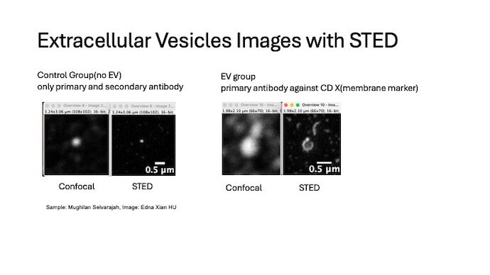
This year, the newly acquired Abberior Facility Line STED/FLIM system was among the hands-on stations. As the most advanced STED/FLIM system in Norway, it bridged resolution capabilities between electron microscopy (EM) and optical microscopy, attracting high-impact projects and extensive bookings.
Workshop participants brought their own samples, gaining preliminary data with this cutting-edge technology in Oslo. A Bergen participant brought Orca primary cells to conduct multiple treatment conditions, while others tested their research samples and explored imaging modalities, gaining insights for future experiments.