Microscope Image Gallery

NRK article about Julien Resseguier's work on fish immunology collected and processed with the system in NorMIC platform(Dragonfly and Imaris Image Processing Workstation).

ECI Cleared 3 day old gastroid (membrane stain). Spinning disk confocal. Diameter across 250 micron. Source: Joachim Mossige, Image: Edna
MDCK Cells Spreading (Red: Focal adhesion protein, Green: Rab 8) - TIRF Microscopy - Source: Edna

Mitochondria of live Hela K cells (STAR Live Orange). (Top: Confocal image, Bottom: STED Image, Scale bar 1 micron) - Source: Andreas Brech, Image: Edna
Mitochondria of live Hela K cells (STAR Live Orange). (Top Left: Confocal overview, Top Right: STED overview, Lower left: Confocal zoom-in, Lower Right: STED zoom-in, Scale bar 5 micron) - Source: Andreas Brech - Image: Edna
Event Photo Gallery
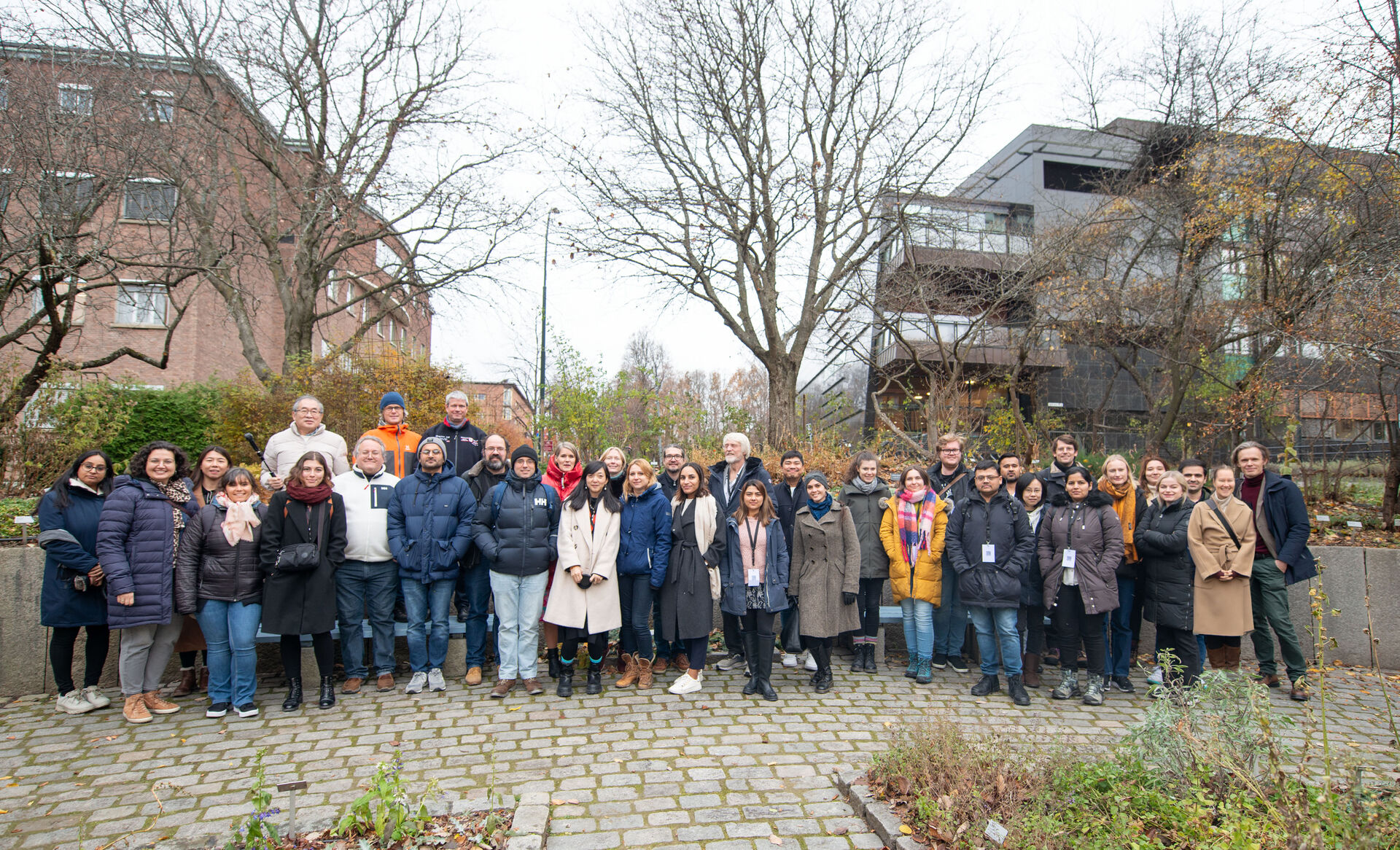
11th NorMIC workshop group photo- 2023 Nov