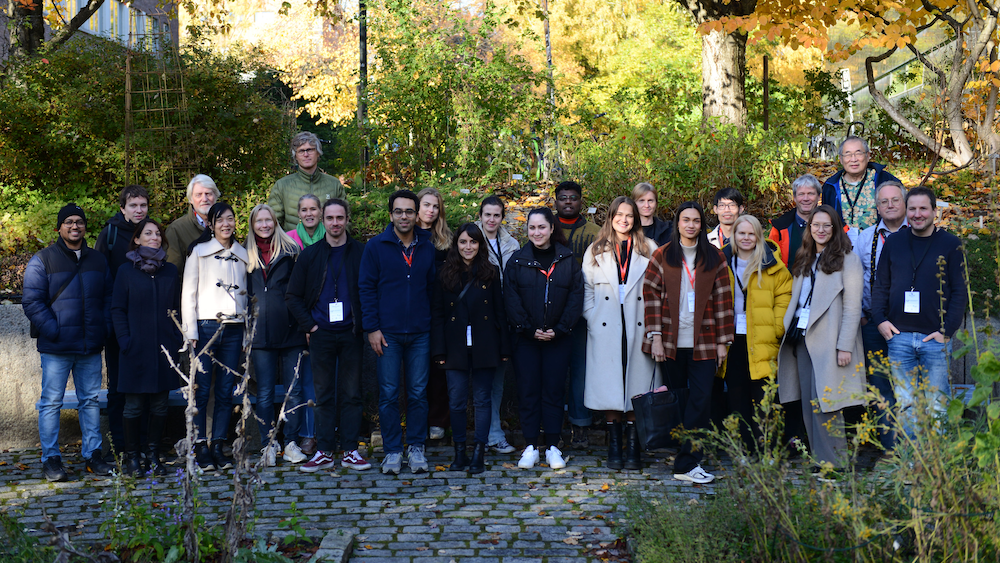
The 12th NorMIC (Norwegian Molecular Imaging Consortium) workshop was successfully held at the Department of Biosciences from October 15th to 18th. This bi-annual event continues to facilitateinvaluable hands-on experience, knowledge sharing, and professional networking among the imaging scientific research community.
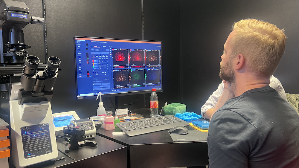
This year's workshop ventured beyond the standard curriculum by introducing novel topics such as FLIM-STED (Fluorescence Lifetime Imaging Microscopy - STimulated Emission Depletion), MINFLUX, and the principles behind Light Sheet Microscopy. These cutting-edge topics have been added because STED-FLIM, MINFLUX and light sheet microscopes have either been purchased already or are in the process of beingacquired by core facilities at the university.
STED and MINFLUX
The NorMIC facility recently welcomed a new member to their instrument family: the Abberior Facility Line STED system, which is now fully operational. Furthermore, the core facility under the leadership of William Louch, is active in the procurement process of a MINFLUX system. By integrating these topics into the workshop, they aim to equip the local user community with the expertise necessary to take full advantage of these novel imaging modalities in their current and upcoming research endeavors.
Light Sheet Microscope
Blindern campus is witnessing a surge in research groups engaged in organoid imaging. A typical organoid easilyexceeds 300 micrometers in diameter, outstripping the working distance of our current high-resolution microscopes. To address the increasing demand for advanced imaging capabilities, the light microscopy facility of the new Life Science building has obtained the funding to acquire a Light Sheet Microscopy system. This addition is set to bridge the existing technological gap in our imaging resources.
The implementation of Light Sheet Microscopy requires specialized knowledge in sample handling and preparation, as well as system operation and maintenance, compared to traditional point scanning or spinning disk confocal microscopes. The variety in commercial systems calls for a customized approach to meet the specific needs of our diverse research community. In anticipation of this new technology, NorMIC conducted a proactive outreach campaign, which included a request for registration from potential users.
Many workshop organizer are also in the procurement committee of the light sheet microscope, they are also relatively new to the light sheet techniques. By involvement in workshop, they have gained significant expertiseas well. Interaction with the students during the workshop has provided them with a deeper understanding of the practical applications and user expectations of light sheet microscopy.
Positive feedback and gratitude
While the workshop's intense agenda is demanding for organizers and participants alike, the spirit of engagement and eagerness to learn has once again defined the experience. The heartfelt and positive feedback received by the organizing committee from attendees is a testament to the workshop's impact and success.
The NorMIC Imaging platform extends its deepest gratitude to all participants, speakers, and its funding agencies (UiO:Life Sciences and Digital Life Norway) for making the NorMIC workshop enriching and enjoyable. Looking ahead, The NorMIC Imaging platform remains committed to supporting the advancement of imaging science and empowering its researchers with the best tools and knowledge.