The Nordic region joins forces
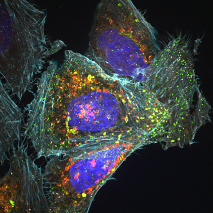
Advanced microscopy gives researchers completely new opportunities to understand how cells and molecules behave, and thus also how disease arises and can be treated. Through the Nordic network “Bridging Nordic Microscopy Infrastructures-II (BNMI-II)”, collaboration between research environments across the Nordic region is being strengthened.
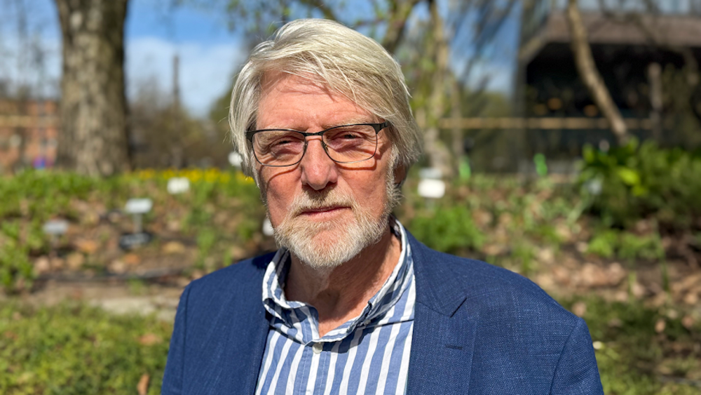
Oddmund Bakke is Professor Emeritus at the Department of Biosciences, University of Oslo. He is the head of BNMI-II and has closely followed developments in the field.
“If someone had told me in the late 1980s, when I was a young researcher working with relatively primitive microscopes, that within a few decades we would be able to study living biological processes at the molecular level with such precision, it would have sounded like science fiction,” Bakke says.
“For many research areas, advanced microscopy has become one of the most important discovery tools in medical science and natural science,” he says.
Advances in imaging technology now make it possible to study proteins in living cells in real time. Previously, cells often had to be chemically fixed, and researchers were limited to static snapshots.
Sharing equipment and expertise
In the laboratories at the University of Oslo, some of the most advanced microscopes in the Nordic region are in place. Researchers from Iceland, among others, come here to learn how to use instruments that will later be moved and put into use there. In this way, both equipment and expertise are shared across the Nordic region.
“It is absolutely crucial that we collaborate in the Nordic region. This is a scientific field developing at a tremendous pace, and when we join forces across the Nordic countries, we can compare ourselves with larger countries such as France, Germany, and Italy,” says Bakke.
“We exchange valuable knowledge with one another, among other things through our annual symposium. This makes working with microscopy much more interesting, and strengthens the research,” he says.
What happens next?
“Microscopy is ultimately limited by the laws of physics, so there are theoretical limits to how many details we can distinguish. At the same time, researchers have repeatedly managed to overcome what were once seen as fundamental barriers. Progress is extremely rapid, and techniques that only a few years ago were considered impossible are now standard. This makes it very difficult to predict the future, but one thing is certain: we will continue to see remarkable advances,” says Oddmund Bakke.
Read the full story on NordForsk’s website: Forsker: Mikroskopet er blandt medicinvidenskabens største opdagelsesværktøjer