
BIO4400: Marine Pelagic Oceanography
In BIO4400, students learn about ocean processes, food webs, species adaptations, and fisheries. Each year, the class ventures out to inner oslofjorden on a research vessel. The field work is completed three times to observe seasonal changes in the pelagic environment from the beginning, middle and end of the Spring semester. I'm not taking this class, but I've heard it is both challenging and rewarding. In addition to the phytoplankton shown below, the class also investigates larger pelagic fish (e.g. cod) and invertebrate species (e.g. shrimp). Not everything is tiny in the ocean, after all.
Here are pictures from their first voyage this Spring.

Shane sampling plankton with a surface tow in inner Oslofjorden.
The little floating critters collect and concentrate in the net
and then can be viewed under a microscope.
Photo: Christopher Hinchcliffe

Professor Bente Edvardsen next to the "Niskin Bottle Rosette",
attached to the CTD (Conductivity-Temperature-Depth Instrument).
This is used to compare water quality from different depths.
Photo: Christopher Hinchcliffe
Fridtjof Nansen is credited with inventing the Nansen bottle, the precursor to the Niskin bottle. See Winter fun and My Background in NH if you would like to read more about Nansen.

Thomas fixing the DO (Dissolved Oxygen) samples.
Photo: Christopher Hinchcliffe

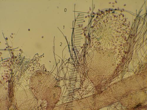
Plankton sample including Diatoms (Coscinodiscus spp.)
Dinoflagellates (Ceratium spp.)
and zooplankton (Calanus spp. & Copepod nauplii).
Photo: Christopher Hinchcliffe

Phytoplankton microscope team!
(Left to right: Istak, Shane, Lars & Petra)
Photo: Christopher Hinchcliffe
BIO4325: Systematics and Ecology of Marine Algae
In BIO4325, students are introduced to the strange and exciting world of algae. Algal species range from bacteria (e.g. Cyanobacteria), responsible for releasing oxygen into the Earth's atmosphere millions of years ago, to giant kelp that extend over 50 meters (e.g. Macrocystis sp.). This is currently my only class and I adore it. Each week we discuss a group of algae (e.g. Stramenopiles) and then go to the microscope lab to view these unusual organisms on slides. You'll notice that the three species shown below do not look very different from a distance, however, they are quite unique from one another on the slides. The reproductive organs are especially useful to tell different species apart (like flowers of angiosperms and cones of gymnosperms).
Here are some pictures from our red algae (Rhodophyta) microscopy lab.
Cells of sporophyte Aglaothamnion hookeri:
The long cells are part of the vegetative plant (seen below),
the round objects are paraspores: one process of asexual reproduction.
Photo: Christopher Hinchcliffe
Aglaothamnion hookeri: Galway, Ireland. 2002.
© Michael Guiry
Carposporophyte cells of Dasya baillouviana on female plant.

The long cells are part of the vegetative plant (seen below),
The carpospore releases carpospores:
a unique step in red algae reproduction.

Photo: Christopher Hinchcliffe

Dasya baillouviana. Schilksee, Kiel Bight, Baltic Sea. 2004.
© Dirk Schories

Spermatangia cells of Dasya sessilis.

The thin cells are part of the vegetative plant (seen below),
The spermatangial branch (cone shaped) has male gametes:
spermatium (male gamete without flagella)

Photo: Christopher Hinchcliffe

Dasya sessilis, Islas Cíes, Galicia, Spain, 2012. © Ignacio Bárbara
MBV4110 - Electron microscopy
This course introduces students to SEM (scanning electron microscope) and TEM (transmission electron microscope) microscopy. Both SEM and TEM use a focused beam of electrons to view specimens. SEM is used to view the outer surface of organisms, while TEM is used to look at cross-sections. The SEM course I took during my undergraduate studies at Colorado College was definitely one of my favorite biology classes. Once you know how to use these microscopes, the possibilities are endless.
Here are some of the photos I took during my SEM course at Colorado College.

Diatom Plankton: Chaetoceros sp. & Achnanthes sp. from the Strait of Juan de Fuca, between US and Canada.
At first I thought these two species were fighting. Turns out, they probably just got tangled in the sample.
Diatoms have siliceous cell walls called frustules. These diatoms are examples of chains of single cells. The Chaetoceros sp. cells have setae protruding from their sides. Setae are believed to aid diatoms with positioning in water currents.

Diatom plankton from the Strait of Juan de Fuca: Eucampia sp.
To me, this diatom's skeleton is art.
The frustule or cell wall is composed of two valves. The valves are covered with pores and areolae. The areolae are hexagonal holes that allow gas and nutrients to diffuse through the frustule.

Pennate diatom residue on eelgrass (Zostera marina). Some diatoms are able to move with a groove known as the raphe. These diatoms can leave slime trails, which allow them to adhere to surfaces.
The boxes in the background are the individual cells of the eelgrass.
Log in to comment
Not UiO or Feide account?
Create a WebID account to comment